Portable 3D scanning has transformed global health initiatives by expanding access to clinical prosthetics, orthotics, and reconstructive care in remote, low-resource regions. In 2022, the World Health Organization and UNICEF estimated that more than 2.5 billion people need at least one assistive product, yet access to these products ranges from 90% of the need in high-income nations to just 3% in low-income ones. Historically, only 5% to 15% of amputees in lower-resource countries have access to prosthetic devices due to prohibitive supply chain costs, lack of trained orthotists, and the logistical burden of traditional plaster casting. By replacing cumbersome plaster casting with lightweight, mobile digital workflows, humanitarian organizations and clinicians overcome long-standing logistical, financial, and geographic barriers.
Proven Deployments in the Field
Several programs demonstrate how digital capture and 3D printing operate in low-resource settings:
- Refugee Camps and Humanitarian Telerehabilitation:
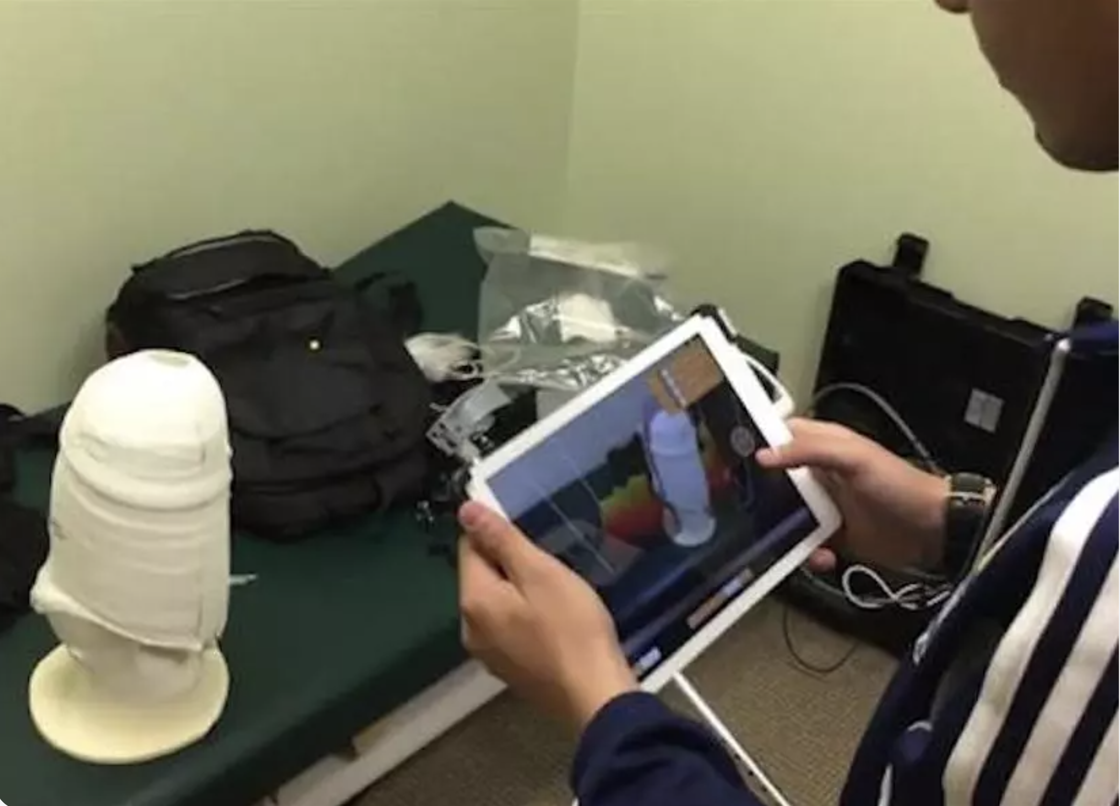 Humanity & Inclusion (HI) introduced handheld 3D scanning and telerehabilitation workflows into refugee settlements across Uganda, Togo, and Madagascar. By mounting compact depth sensors on tablet computers, field clinicians digitize limb anatomies in remote camps in under 60 seconds. This eliminated the need to transport hundreds of pounds of plaster or send vulnerable patients across long, unsafe travel routes for manual limb impressions.
- Community Outreach and Field Clinics: In the Petén region of Guatemala, organizations like LifeNabled partner with local providers like Hospital Shalom to operate biannual clinics delivering comprehensive prosthetic care. Multidisciplinary teams—comprising certified prosthetists, technicians, medical staff, and volunteer support—evaluate, fabricate, and fit devices for dozens of patients per trip at zero cost to them or their families, bridging the gap between specialized clinical care and remote, low-resource populations.
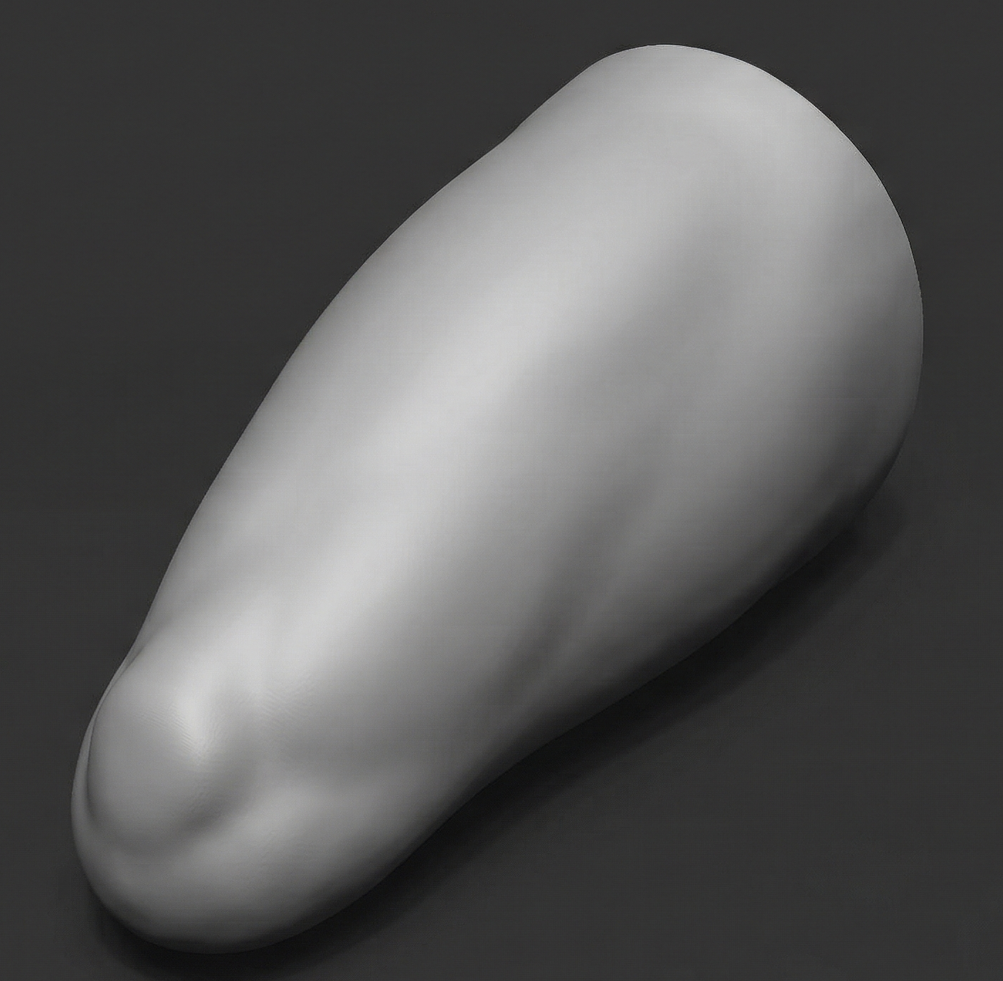
- Conflict Zones and Post-War Rehabilitation: Through initiatives like the Victoria Hand Project,portable 3D scanners have been deployed in Guatemala to capture socket geometries for war-wounded patients and children with congenital limb differences without requiring physical contact on sensitive or scarred post-surgical tissue. In remote regions of Armenia, healthcare workers use portable scanners connected to cloud platforms to digitize the limbs of war victims locally, allowing off-site prosthetists to design custom socket fits remotely.
What the Evidence Says
Systematic reviews and clinical evaluations show that modern tablet- and smartphone-compatible 3D depth sensors achieve high geometric accuracy for lower- and upper-limb socket fitting. They significantly outperform legacy casting methods in speed, patient preference, and portability.
What Scanning Will Not Fix
Where clinicians, printers, materials, and follow-up care are scarce, a better scan will not magically solve all problems. Any program should plan for:
- Training and Fabrication: Someone has to know what a good scan looks like and how to turn it into a well-fitting device. Additionally, a scan has to reach a printer or workshop that can produce a durable device.
- Validation and Limits: A scanner captures shape, but it does not diagnose or prescribe. Like most optical scanners, it can also struggle with very dark or highly reflective surfaces. As the Cambodia study shows, test any scanner against an experienced clinician before relying on it.
Where the Structure Platform Fits
The Structure platform's architecture is uniquely suited to offer solutions for remote, resource strapped environments:
- Offline Operation: Structure Capture scans without an internet connection, which is a requirement where connectivity is unreliable.
- Optimized Scanning Modes: Structure helps lift the training burden by offering preset scanning modes for different scanning environments, including prosthetic sockets and lower extremities.
- Field Durability: Structure Sensor 3 offers 3.5+ hours of continuous battery life and can scan outdoors in direct sunlight, which suits places without a dedicated scanning room.
- Accessible Hardware and Open Formats: While optimized for the Structure Sensor 3, Structure Capture runs on an iPhone or iPad utilizing Apple's TrueDepth camera array, keeping costs down while still delivering scanning needs. Scans export as STL, OBJ, or PLY to work with whatever CAD and printing tools a program already uses.
- Guided Capture: A real-time quality indicator and built-in landmark detections reduce the experience needed to get a usable scan, which matters where trained staff are scarce.
A Practical Way to Start For a clinic exploring prosthetic design from 3D scans, a small pilot is a great first step:
- Pick one device type, such as an AFO or a foot orthosis.
- Scan a small group of patients and, in parallel, take the usual cast or measurement.
- Compare repeatability and fit, with an experienced clinician judging the result.
- Decide whether the scanning step saves enough time and cost to justify scaling up.
If you run or fund a program in a low-resource setting, contact the Structure team to discuss a pilot. If you are looking to read more about how 3D scanning is personalizing healthcare, check out this article here: Custom Orthotics & Prosthetics: How 3D Scanning Is Personalizing Healthcare.
References
- Abbady, H. E. M. A., Klinkenberg, E. T. M., de Moel, L., et al. (2021). 3D-printed prostheses in developing countries: A systematic review. Prosthetics & Orthotics International, 46(1), 19–30.
- Cabibihan, J.-J., Alkhatib, F., Mudassir, M., et al. (2021). Suitability of the Openly Accessible 3D Printed Prosthetic Hands for War-Wounded Children. Frontiers in Robotics and AI, 7.
- Dickinson, A., Donovan-Hall, M., Kheng, S., et al. (2020). Selecting Appropriate 3D Scanning Technologies for Prosthetic Socket Design and Transtibial Residual Limb Shape Characterisation.
- Nia Technologies, University of Toronto, March 2019.
- Prothea, MIT Solve, updated Feb 2024.
- Silva, R., Silva, B., Fernandes, C., et al. (2024). A Review on 3D Scanners Studies for Producing Customized Orthoses. Sensors, 24(5), 1373.
- Ugani Prosthetics, Zero Project, Dec 2023.
- University of Southampton / Exceed Worldwide Cambodia study, Journal of Prosthetics and Orthotics, 2021.
- van der Stelt, M., Verhulst, A. C., Vas Nunes, J. H., et al. (2020). Improving Lives in Three Dimensions: The Feasibility of 3D Printing for Creating Personalized Medical Aids in a Rural Area of Sierra Leone. The American Journal of Tropical Medicine and Hygiene, 102(4), 905–909.
- WHO/UNICEF news release, 16 May 2022.